
2D ECHO
An echocardiogram (echo) is a graphic outline of the heart's movement or is a test which uses sound waves to produce live images of your heart. During an echo test, ultrasound (high-frequency sound waves) from a hand-held wand placed on your chest provides pictures of the heart's valves and chambers and helps the sonographer evaluate the pumping action of the heart. In short it monitors the functioning of the heart and its valves to determine the health of heart muscle especially after the heart attack, can also reveal defects in heart in unborn babies.
The images can help in spotting :
• Blood clots in the heart
• Fluid in the sac around the heart
• Problems in the aorta, which is the main artery connected to the heart.
A doctor advices echocardiogram for a couple of reasons mostly:
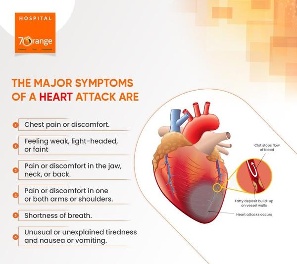
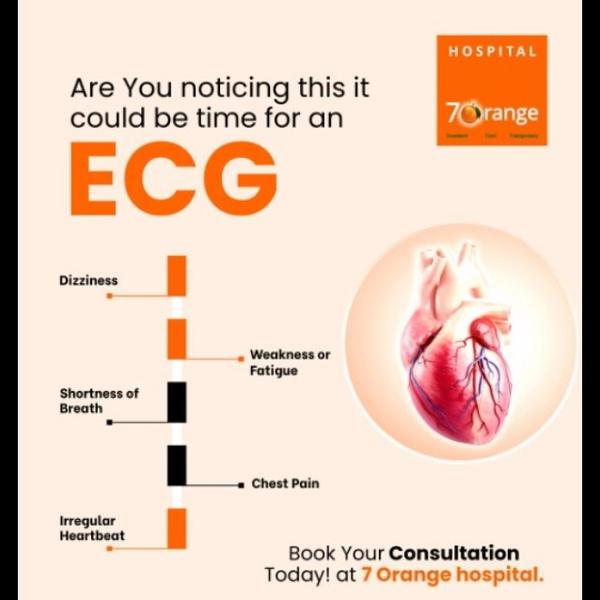
If you have an irregular heartbeat, your doctor may want to inspect the heart valves or chambers or check your heart’s ability to pump. They may also order one if you’re showing signs of heart problems, such as chest pain or shortness of breath.
There are several different types of echocardiograms :
1. Transthoracic echocardiography
2. Transesophageal echocardiography
3. Stress echocardiogram
4. Three-dimensional echocardiography
5. Fetal echocardiograph
Transthoracic Echocardiography : This is the most common type of echocardiography which is painless and non-invasive. A device called a transducer will be placed on your chest over your heart. During the test you will be asked to lie flat on your back as wires are placed on your chest to monitor your heart rhythm. The sonographer will instruct you to turn on your left side and the probe will be placed on your chest. A lubricant (gel) is used to improve picture quality and this may feel cold. Gentle pressure is applied onto your chest as necessary in order to obtain the information but generally should not be painful. Let the sonographer know if he or she is pressing too hard. You will see images of your heart on the monitor, some of which will have color. The colour and the sounds that you may hear coming from the machine are representations of blood flow within your heart.
Transesophageal echocardiography : If a transthoracic echocardiogram doesn’t produce definitive images, your doctor may recommend a transesophageal echocardiogram. In this procedure, the doctor guides a much smaller transducer down your throat through a thin, flexible tube in your mouth. They will numb your throat to make this procedure easier. This tube is guided through your esophagus, the tube that connects your throat to your stomach. With the transducer behind your heart, your doctor can get a better view of any problems. If you undergo a transesophageal echocardiogram, your doctor may instruct you not to eat anything for a few hours before the test. This is to prevent you from vomiting during the test. You may also not be able to drive for a few hours afterward due to the sedatives.
Stress echocardiogram : A stress echocardiogram uses traditional transthoracic echocardiography. However, the procedure is done after you’ve exercised or taken medication to make your heart beat faster. This allows your doctor to test how your heart performs under stress. One must wear clothes and shoes that are comfortable to exercise in.
Three-dimensional echocardiography : A three-dimensional (3-D) echocardiogram uses either transesophageal or transthoracic echocardiography to create a 3-D image of your heart. This involves multiple images from different angles. It’s used prior to heart valve surgery. It’s also used to diagnose heart problems in children.
Fetal echocardiography : Fetal echocardiography is used on expectant mothers sometime during weeks 18 to 22 of pregnancy. The transducer is placed over the woman’s belly to check for heart problems in the fetus. The test is considered safe for an unborn child because it doesn’t use radiation, unlike an X-ray.
Echocardiograms are considered very safe. Unlike other imaging techniques, such as X-rays, echocardiograms don’t use radiation. Your doctor will review your results after the test. The results may reveal abnormalities such as:
• damage to the heart muscle
• heart defects
• heart size
• pumping strength
• valve problems
If your doctor is concerned about your results, they may refer you to a cardiologist. This is a doctor who specializes in the heart. Your doctor may order more tests or physical exams before diagnosing you. If you’re diagnosed with a heart condition, your doctor will work with you to develop a treatment plan that works best for you.
Note: At 7 Orange Hospital we conduct “Transthoracic Echocardiography for only Rs. 1000/- from Monday to Saturday. To book an appointment kindly call us @ 7350055754.