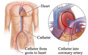
Coronary catheterization :-
A coronary angiogram (an X-ray with radiocontrast agent in the coronary arteries) that shows the left coronary circulation. The distal left main coronary artery (LMCA) is in the left upper quadrant of the image. Its main branches (also visible) are the left circumflex artery (LCX), which courses top-to-bottom initially and then toward the centre/bottom, and the left anterior descending (LAD) artery, which courses from left-to-right on the image and then down the middle of the image to project underneath the distal LCX. The LAD, as is usual, has two large diagonal branches, which arise at the centre-top of the image and course toward the centre/right of the image.
Indications for cardiac catheterization include the following:
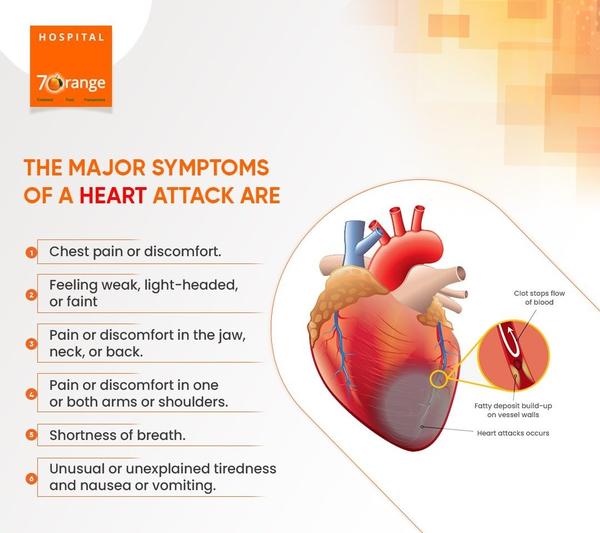
• Heart Attack (includes ST elevation MI, Non-ST Elevation MI, Unstable Angina)
• Abnormal Stress Test
• New-onset unexplained heart failure
• Survival of sudden cardiac death or dangerous cardiac arrhythmia
• Persistent chest pain despite optimal medical therapy
• Workup of suspected Prinzmetal Angina (coronary vasospasm)[1]
Coronary catheterization is performed in a catheterization lab, usually located within a hospital. With current designs, the patient must lie relatively flat on a narrow, minimally padded, radiolucent (transparent to X-ray) table.
The X-ray source and imaging camera equipment are on opposite sides of the patient's chest and freely move, under motorized control, around the patient's chest so images can be taken quickly from multiple angles. More advanced equipment, termed a bi-plane cath lab, uses two sets of X-ray source and imaging cameras, each free to move independently, which allows two sets of images to be taken with each injection of radiocontrast agent.