CARDIAC CATHERISATION :
OUR APPROACH:
Cardiac Catherisation provides us information of your Hearts blood vessels and pressures inside it. This procedure is done to achieve maximum accuracy in diagnosis of heart related symptoms like fatigue, breathlessness or short breath, dizziness, chest pain – Angina.
We at 7 Orange Hospital, Chinchwad perform more than 100 catherisations every year. Our sophisticated Cath Lab uses low-radiation, high resolution equipments to maximise patient safety along with the image quality. Our radial technique (through the wrist) improves comfort level of the patient thereby reducing the recovery time.
Overview of Cardiac Catherisation :
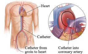
This procedure is used to diagnose different types of heart diseases and its varying conditions. At the time of procedure our highly trained and skilled Interventional Cardiologist either places a sheath (small tube like an IV) through the skin into an artery via Wrist OR Groin. They pass catheter towards the heart of the patient. These are called as minimally invasive procedures which helps to diagnose and treat versatile range of problems with your heart’s chambers, valves and vessels.
Various Conditions treated via Cardiac
Catherisation :
We advice Cardiac Catherisation to those patients who experience chest pain or breathlessness, dizziness or fatigue. We use cardiac catherisation for various conditions like :
1. Cardiomyopathy (Angina/ Heart attack)
2. Congenital heart disease, including atrial septal defects and Ventricular septal defects.
3. Coronary Artery Disease (CAD)
4. Heart failure
5. Valvular Heart disease.
6. Rhythm disturbances (either too fast or too slow heart rate)
Types of Cardiac Catherisation : During Cardiac Catherisation we perform such as :
• Coronary Angiogram or Angiography: In this procedure blocked arteries which are associated with heart diseases are located. Cardiologist injects a special contrast dye that shows up on low dose X-rays and tracks the flow of blood. Cardiologist can locate blocked arteries, assess the severity of the condition and determine what type of treatment is required.
• Fractional Flow reserve : In Angiogram if an artery is modestly narrowed then this test helps to indentify the need for treatment. We thread a wire with a pressure sensor past the spot in question, to compare blood flow and pressure on each side. We can identify when you require a stent and when medication alone is enough.
• Intravascular Ultrasound (IVUS) : An ultrasound probe to the end of Cathether is attached to observe inside arteries and to make measurements incase, vessels are narrowed or blocked. This technique often provides more information than a standard angiogram and can help to schedule appropriate treatment. We can also evaluate suspect bridging when an artery sits under heart muscle rather than on top. Thus, IVUS technique is highly precise tool for detecting and evaluating any Coronary artery disease.
• Optical Coherence Tomography (OCT) : In this procedure a Probe is used like IVUS, OCT uses a probe but one that emits light waves instead of sound waves. The resolution of OCT is higher as compared to IVUS, with the ability to see tiny details inside an artery.
• DSA for visualisation of peripheral arteries.
• 4 vessel angiography for pt. Having CVA (Stroke)
• Renal Angiography : For patients whose BP is not controlled by medications.
Before the procedure:
Briefing about the procedure is done to the patient and consent form is also filled by patient to give permission to proceed with the procedure. Patient is kept on NBM (Nil by mouth OR nothing by mouth) i.e, on fast for overnight before the procedure. Prior to procedure routine blood test is done to check blood clotting time. Other blood tests may be done as well. Based upon certain medical conditions, doctor may request other specific preparation.
During the Procedure :
A hospital gown will be provided to you for wearing. Nurse will insert an IV i.e. Intravenous line in your arm in order to provide medication and fluids. Local Anaesthesia is given to the arm to avoid pain at the insertion site. Sheath is inserted into the blood vessel in your groin or arm wrist – radial approach. Through this sheath a thin long tube called catheter is placed inside the artery. This catheter is properly guided towards the heart to perform various tests, to check other parts of heart. Sterile drapes are used to cover the site to help prevent any infection. For proper visualisation of artereies a contrast dye is injected through the catheter.
After the Procedure :
The Cathlab technician informs the patient how long to lie down and keep the insertion site still. If the insertion is done through groin region then patient is asked to stay keeping his legs in same position for more than 2 hours. On the artery site if closure device like collagen plug or Suture is used then the patient may be able to move sooner however, this totally depends on how much bleeding occurs on the site during insertion. Nurse will monitor your blood pressure and insertion site. If required the patient may be asked to drink some fluid to flush the contrast liquid out of the system. A small lump or bruise swelling will occur at insertion site which goes away in few weeks and this is almost normal to all patients. Patient is usually discharged on same day following diagnostic catherisation after 4-6 hours of observation.
We are known as “Centre for Excellence in Cardiology”. We have expert team of Cardiologist, Cardiac Surgeons, and Cardiac Anaesthetist who perform various cardiac procedures, available 24 X 7. We also provide Intensivists who handles critical cases with excessive care. For economically backward patients we are authorised with “Chief Minister Relief Fund” to benefit Angiography at Rs. 9999/- and Angioplasty at Rs. 99999/- . For further enquires please feel free to call us at 7350055754 – 7 Orange Hospital. Pawana nagar, Chapekar chowk, Chinchwad, Pune – 411033.